
Evaluation of Anti-inflammatory Efficacy via TNF-α in an Ex-Vivo Model using the ELISA Technique
An ex-vivo human skin model consists of human skin explants obtained through ethically approved procedures, often from cosmetic surgeries such as abdominoplasties or mammoplasties, and subsequently cultured under controlled laboratory conditions, including specific media, temperature, and humidity, to maintain tissue viability and functionality. This model is of paramount importance in dermatological and cosmetic research, particularly for evaluating the efficacy of various active substances, including those with anti-inflammatory properties, thereby offering a significant advantage over simplistic 2D cell cultures or less directly translatable animal models. A key advantage of the ex-vivo model is the preservation of the human skin’s native architecture.
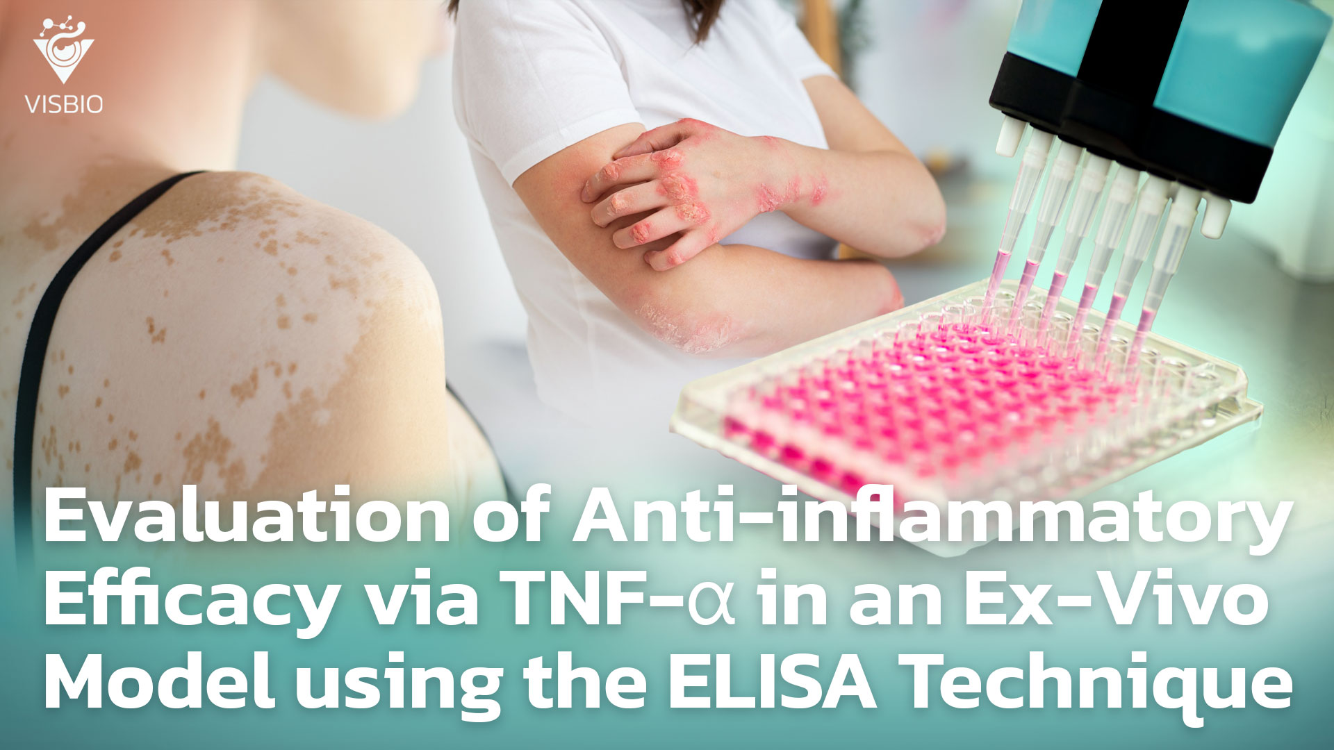
In testing anti-inflammatory activity, Tumor Necrosis Factor-alpha (TNF-α), a potent pro-inflammatory cytokine, plays a crucial role in stimulating and promoting inflammatory processes in the skin. Therefore, measuring TNF-α levels serves as an important indicator for assessing the efficacy of anti-inflammatory agents. The Enzyme-Linked Immunosorbent Assay (ELISA) technique is a widely adopted method for quantifying TNF-α in samples or lysates from ex-vivo skin models. This technique relies on specific antigen-antibody interactions and an enzymatic reaction that produces a measurable signal, typically a color change, proportional to the amount of TNF-α present. The trend towards reducing animal testing and the development of New Alternative Methods (NAMs) further underscore the increasing importance of ex-vivo human skin models in the pharmaceutical and cosmetic industries. These NAMs are crucial for meeting regulatory demands and societal expectations for more humane and scientifically robust testing strategies, as they represent ethical testing methods that provide directly relevant human data.
Tumor Necrosis Factor-Alpha (TNF-α): Skin Inflammation
Tumor Necrosis Factor-alpha (TNF-α) is a cytokine that plays a critical role in mediating and promoting inflammatory processes within the skin. TNF-α is produced by various cell types in the skin, including immune cells such as macrophages, T lymphocytes, and mast cells, as well as keratinocytes, the primary cells of the epidermis. When the skin encounters stimuli, such as pathogen-associated molecular patterns (PAMPs) from microbes, damage-associated molecular patterns (DAMPs) from injured cells, or other inflammatory signals, these cells secrete TNF-α to initiate and regulate immune and inflammatory responses. TNF-α exerts its effects by binding to specific cell surface receptors, primarily TNFR1 and TNFR2, which trigger distinct intracellular signaling pathways often culminating in the activation of transcription factors like NF-κB, a key regulator of inflammatory gene expression.
Role of TNF-α in Promoting Inflammation
TNF-α performs several functions to promote inflammation in the skin:
- Activation of Immune Cells: TNF-α activates immune cells, such as neutrophils and macrophages, inducing their migration to the site of inflammation to eliminate foreign agents or damaged cells through processes like phagocytosis and the release of antimicrobial substances or reactive oxygen species.
- Production of Other Inflammatory Mediators: TNF-α stimulates skin cells and immune cells to secrete other cytokines and chemokines involved in inflammation, such as Interleukin-1 (IL-1) and Interleukin-6 (IL-6), thereby amplifying the inflammatory cascade. These mediators often act synergistically, creating a complex and self-perpetuating inflammatory milieu.
- Increased Expression of Adhesion Molecules: TNF-α stimulates endothelial cells to increase the expression of adhesion molecules, such as ICAM-1 and VCAM-1, which facilitates the adhesion of immune cells to the vascular endothelium and their subsequent infiltration into inflamed skin tissue.
- Stimulation of Keratinocytes: TNF-α can stimulate keratinocytes to proliferate and secrete inflammatory mediators, a characteristic observed in certain inflammatory skin diseases, such as psoriasis. This can also contribute to epidermal hyperplasia and potentially compromise the skin’s barrier integrity.
Relevance of TNF-α to Inflammatory Skin Diseases
Dysregulation in the production or function of TNF-α is linked to the onset and progression of several inflammatory skin diseases, such as:
- Psoriasis: Elevated levels of TNF-α in the skin of psoriasis patients play a significant role in stimulating inflammation, abnormal keratinocyte proliferation, and angiogenesis.
- Atopic Dermatitis: TNF-α is involved in the aberrant immune responses and skin barrier dysfunction in patients with this condition.
- Hidradenitis Suppurativa (HS): TNF-α is a key cytokine implicated in the chronic inflammation characteristic of HS.
- Other Skin Diseases: Including Vitiligo and skin damage resulting from ultraviolet (UV) radiation exposure.
Due to its significant and broad role in inducing inflammation, TNF-α serves as both an important biomarker for assessing the inflammatory state of the skin and a primary target for the development of many anti-inflammatory drugs. The selection of TNF-α as an indicator for testing the efficacy of anti-inflammatory substances in ex-vivo models is highly rational, given its direct link to inflammatory mechanisms in the skin.
Ex-Vivo Human Skin Model: Assessing Anti-inflammatory Efficacy by Measuring TNF-α with the ELISA Technique
The ex-vivo human skin model is an exceptionally valuable tool in dermatological research, particularly for evaluating the anti-inflammatory effects of active substances or products. This is due to its close resemblance to the natural physiology of human skin, which includes multiple interacting cell types, a three-dimensional tissue structure, and functional barrier properties, while still maintaining the complexity of the skin’s overall architecture. Furthermore, the use of ex-vivo human skin models helps to avoid ethical concerns associated with animal testing, as it is well-known that animal skin can differ significantly from human skin in terms of microanatomy, immunology, and physiology, making data extrapolation challenging.
The ability to use test substances in an ex-vivo model allows for the precise evaluation of compound penetration and the stimulation or inhibition of specific biological responses within the human tissue matrix. Specifically, changes in the levels of Tumor Necrosis Factor-alpha (TNF-α), a key cytokine involved in inflammation, can be measured using the specific and accurate ELISA technique. The measurement of TNF-α in this ex-vivo model, which closely mimics clinical application scenarios by allowing topical application of formulations to the skin surface, enables a reliable assessment of the efficacy of substances aimed at reducing inflammation directly within a relevant human tissue context. This provides more robust and translatable data for predicting clinical outcomes compared to simpler in-vitro systems.
Evaluation of Anti-inflammatory Efficacy (TNF-α) in Ex-Vivo Models using ELISA: Suitable Examples
Inflammation is often caused by external factors such as UV radiation, heat, humidity, pollution, or irritation, leading to symptoms like redness, swelling, and heat. TNF-α (Tumor Necrosis Factor-alpha) is a protein belonging to the cytokine group that plays a critically important role in initiating and promoting the inflammatory process in the body, including in the skin. TNF-α is a key mediator that triggers various inflammatory signals, stimulates immune cell activity, and leads to the subsequent release of other pro-inflammatory substances. Elevated levels of TNF-α are an indicator of inflammation occurring in the skin.
The evaluation of anti-inflammatory efficacy using TNF-α as a marker in Ex-Vivo models involves applying the product or substance to be tested to Ex-Vivo skin samples, then inducing inflammation, and subsequently measuring the quantity of TNF-α produced by the skin cells in those samples using the ELISA technique, which is a specific and accurate method for protein quantification. If a product can reduce the elevated levels of TNF-α resulting from inflammation, it indicates that the product likely possesses anti-inflammatory properties.
Examples of suitable products, active ingredients, and topical medicines for this testing include:
1.Products:
- Skincare products for sensitive/easily inflamed skin: Such as moisturizers, serums with soothing and redness-reducing properties.
- Products to reduce redness from acne: As TNF-α plays a role in acne inflammation.
- Products for skin recovery after laser or treatments: Helps reduce inflammation caused by procedures.
2.Active Ingredients:
- Plant or herbal extracts with high anti-inflammatory activity: Such as Curcuminoids from turmeric, Flavonoids, green tea extract, Boswellia serrata extract.
- Synthetic compounds that inhibit TNF-α.
- Substances with antioxidant properties.
3.Topical Medicine
Literature:
- Pocino, K., Carnazzo, V., Stefanile, A., Basile, V., Guerriero, C., Marino, M., Rigante, D., & Basile, U. (2024). Tumor Necrosis Factor-Alpha: Ally and Enemy in Protean Cutaneous Sceneries. International Journal of Molecular Sciences, 25(14), 7762.
- Vugler, A., O’Connell, J., Nguyen, M. A., Weitz, D., Leeuw, T., Hickford, E., Verbitsky, A., Ying, X., Rehberg, M., Carrington, B., Merriman, M., Moss, A., Nicholas, J.-M., Stanley, P., Wright, S., Bourne, T., Foricher, Y., Zhu, Z., Brookings, D., Horsley, H., Heer, J., Schio, L., Herrmann, M., Rao, S., Kohlmann, M., & Florian, P. (2022). An orally available small molecule that targets soluble TNF to deliver anti-TNF biologic-like efficacy in rheumatoid arthritis. Frontiers in Pharmacology, 13, 1037983.