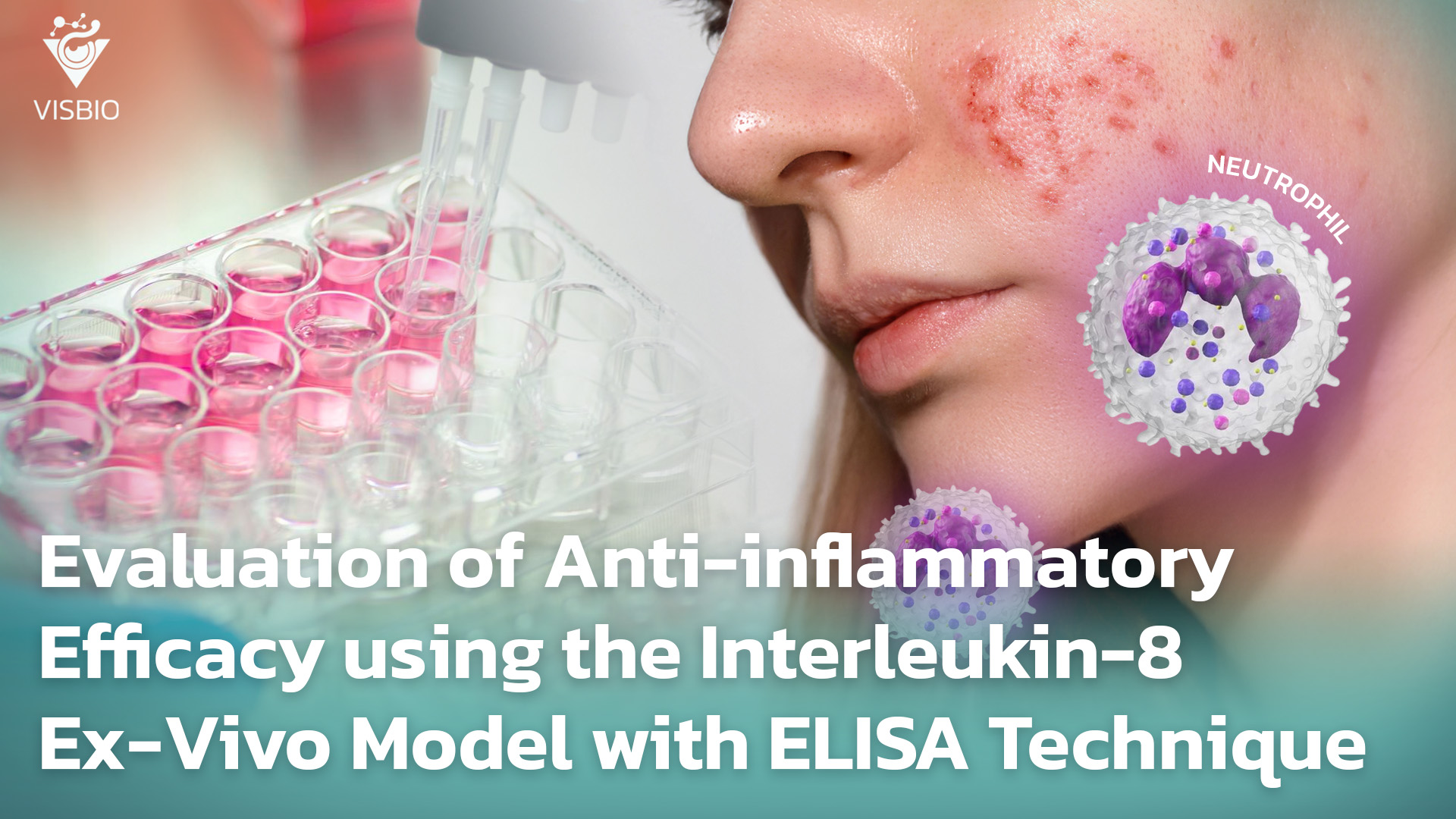
การประเมินประสิทธิภาพการต้านการอักเสบ Anti-Inflammatory - IL 8 (Interleukin-8) แบบจำลอง Ex-Vivo ด้วยเทคนิค ELISA
Skin inflammatory conditions represent a significant challenge directly impacting both the health and satisfaction of consumers. The research and development of active ingredients possessing genuine anti-inflammatory properties, supported by robust scientific evidence, is thus paramount for manufacturers and brand owners aiming to meet market demands and build product credibility.
Interleukin-8 (IL-8) is a key chemokine playing a prominent role in the initiation and persistence of the inflammatory process in the skin. IL-8 levels are frequently used as an indicator of inflammatory status, as they can accurately reflect the impact of various stimulating factors, including the efficacy of substances with anti-inflammatory effects. Therefore, evaluating changes in IL-8 levels is a highly valuable approach.
Role of Interleukin-8 (IL-8) in Skin Inflammation and Wound Healing
IL-8 is a potent chemoattractant for neutrophils, guiding these cells to areas of inflammation, infection, or injury. Furthermore, IL-8 stimulates neutrophil functions such as phagocytosis, degranulation (releasing antimicrobial proteins and enzymes), and respiratory burst (producing reactive oxygen species – ROS). IL-8 is also known to promote angiogenesis, which is relevant in both inflammation and wound healing.
- Role in Wound Healing: In the early phase of inflammation, IL-8 assists in the recruitment of leukocytes to clear cellular debris. Additionally, IL-8 can stimulate the migration and proliferation of keratinocytes, which are essential for re-epithelialization. Studies have found that “rhIL-8 significantly increases keratinocyte migration.” The dual role of IL-8, both in promoting acute inflammation (neutrophil recruitment) and participating in tissue repair processes (angiogenesis, keratinocyte migration), poses challenges in treatment. Effective anti-inflammatory strategies targeting IL-8 must focus on modulation rather than complete inhibition, to avoid disrupting essential wound healing mechanisms. This subtle difference is crucial for product development.
Cellular Sources and Regulation of IL-8 Production in Skin
Various skin cells and immune cells can produce IL-8 when stimulated:
- Keratinocytes: Are a major source of IL-8 in the epidermis, producing IL-8 in response to various stimuli such as PAMPs (e.g., LPS via TLRs), pro-inflammatory cytokines, and tissue injury. It has been noted that “Keratinocytes are a rich source of IL-8” and described that keratinocytes produce IL-8 in response to TLR stimulation. This demonstrates that keratinocytes are not merely passive barrier cells but active players in immune responses.
- Fibroblasts: Fibroblasts in the dermis also contribute to IL-8 production, particularly in response to inflammatory signals.
- Resident Immune Cells: Macrophages, mast cells, and endothelial cells within the skin can produce IL-8.
- Regulation of Production: The expression of IL-8 is strictly controlled and rapidly induced by inflammatory stimuli. Factors such as ROS can also induce IL-8 production.
IL-8 as a Biomarker for Evaluating Anti-inflammatory Product Efficacy
The significant role of IL-8 in initiating and amplifying acute inflammation (neutrophil recruitment) makes IL-8 a primary target for interventions aimed at reducing redness, swelling, and irritation. The involvement of IL-8 in conditions such as psoriasis and irritant responses further confirms the utility of IL-8 as an indicator of product efficacy.
- Case Studies from Research: Research studies have demonstrated the reduction of IL-8 by various test substances/formulations in ex-vivo human skin models, such as Dexamethasone, Betamethasone, formulations for sun damage that reduce UV-induced IL-8, and extracts from natural products or other active ingredients. The ability to induce specific types of inflammation in the ex-vivo model and then measure the reduction of IL-8 allows for testing products against relevant factors, making the efficacy data more applicable to real-world scenarios where skin faces these specific challenges. This significantly strengthens the credibility of product claims.
Ex-Vivo Human Skin Model: A Realistic Platform for Evaluating Anti-inflammatory Effects by Measuring Interleukin-8 (IL-8) using the ELISA Technique
The Ex-Vivo human skin model is a highly valuable tool in dermatological research, particularly for evaluating the anti-inflammatory efficacy of active ingredients or products. This is due to its close resemblance to the physiology of natural human skin, while maintaining the complexity of the skin structure.
Furthermore, the use of ex-vivo human skin models helps avoid ethical concerns associated with animal experimentation. Animal skin is known to differ significantly from human skin in terms of microanatomy and physiology.
The ability to apply test substances in the ex-vivo model allows for accurate evaluation of compound penetration and the modulation (stimulation or inhibition) of localized biological responses within the human tissue matrix. Specifically, changes in the levels of Interleukin-8 (IL-8), a key cytokine involved in inflammation, can be measured using the specific and precise ELISA technique. Measuring IL-8 levels in this ex-vivo model, which closely simulates clinical application scenarios, facilitates reliable evaluation of the efficacy of substances aimed at reducing inflammation.
Therefore, the ex-vivo model is highly suitable for evaluating the efficacy of substances possessing anti-inflammatory properties through the measurement of inflammatory markers such as IL-8 using the ELISA technique.
Evaluation of Interleukin-8 Anti-inflammatory Efficacy in Ex-Vivo Models using ELISA: Suitable Examples
Interleukin-8 plays a significant role in several skin diseases characterized by neutrophil accumulation, such as acne (especially inflammatory acne), psoriasis, and certain types of dermatitis. Evaluating anti-inflammatory efficacy by measuring IL-8 levels in Ex-Vivo human skin models using the ELISA technique is a method used to assess whether a product or substance can inhibit the secretion of IL-8. If a product can reduce the increase in IL-8 levels when the skin is stimulated to induce inflammation, it indicates that the product has the effect of reducing the recruitment of neutrophils to the inflamed area, which is a mechanism of anti-inflammation.
Examples of suitable products, active ingredients, and topical medicines for this testing include:
1. Products:
- Products for the treatment of inflammatory acne.
- Products to soothe redness and swelling from acute inflammation: Such as from insect bites or severe irritation.
- Skincare products for skin diseases where neutrophils are a key mechanism: Such as psoriasis.
- Products used after procedures.
2. Active Ingredients:
- Plant extracts/natural substances with chemokine secretion-reducing activity: Such as extracts known to affect neutrophil migration.
- Anti-inflammatory substances that inhibit IL-8 production pathways.
- Substances with general anti-inflammatory properties: Such as Centella asiatica extract (some studies have shown effects on IL-8), cannabinoid compounds.
- Peptides or molecules that inhibit IL-8 Receptor function.
3.Topical Medicine:
- Topical medications for acne with anti-inflammatory activity: Such as Clindamycin (also has anti-inflammatory activity), Benzoyl Peroxide (some mechanisms involve bacterial reduction and effects on inflammation).
- Topical medications for psoriasis: Which often have mechanisms that reduce inflammation and cell recruitment.
- Topical medications used in the management of inflamed wounds.
Literature:
- Moneo-Sánchez, M., de Pablo, N., Arana-Pascual, L., Beitia, I., Benito-Cid, S., & Pérez-González, R. (2024). Multifunctional, Novel Formulation for Repairing Photoaged and Sun-Damaged Skin: Insights from In Vitro, Ex Vivo, and In Vivo Studies. Cosmetics, 11(6), 224.
- Petrinić Grba, A., Štefok, D., Cedilak, M., Paravić Radičević, A., Antolić, M., Ogrljenović, A., Vidović Iviš, S., Milutinović, V., Čuzić, S., Bosnar, M., & Eraković Haber, V. Human skin explant as a model for inflammation and wound healing. Selvita.
- Shkundin, A., & Halaris, A. (2024). IL-8 (CXCL8) Correlations with Psychoneuroimmunological Processes and Neuropsychiatric Conditions. Journal of Personalized Medicine, 14(5), 488.
- Wong, R. S.-Y., Tan, T., Pang, A. S.-R., & Srinivasan, D. K. (2025). The role of cytokines in wound healing: from mechanistic insights to therapeutic applications. Exploratory Immunology, 2025(5), 1003183.

