

About Our Anti-Skin Cancer Testing Service
VISBIO Co., Ltd. offers laboratory testing of anti-skin cancer activity against human epidermoid carcinoma (A431) cells, carried out by scientific experts. The cancer cells are grown in two-dimensional culture (2D cell culture). This test is suited to in-depth research aimed at developing herbs that can inhibit cancer cells.
Skin cancer is a malignant tumor caused by the abnormal growth and division of the cells that make up the skin, such as epidermal cells, hair follicle cells, sebaceous glands, sweat glands and pigment-producing cells (melanocytes); it can also be cancer that has spread from other organs. The most common skin cancer is basal cell carcinoma, which arises from cells in the basal layer of the epidermis and the skin appendages, followed by squamous cell carcinoma, which arises from cells in the epidermis. Malignant melanoma, which arises from pigment-producing cells, is less common but is the most aggressive and can be fatal. Epidermoid carcinoma, studied using the human A431 cell line, is a type of non-melanoma skin cancer that is less aggressive and has a lower mortality rate than malignant melanoma. Many studies have used A431 human epidermoid carcinoma cells to test inhibitory activity against skin cancer cells in vitro, in microplates (well plates) in which the cancer cells are grown in two-dimensional culture (2D cell culture) in culture medium.
Understanding Skin Cancer
Skin cancer is a malignant tumor caused by the abnormal growth and division of the cells that make up the skin, such as epidermal cells, hair follicle cells, sebaceous glands, sweat glands and pigment-producing cells (melanocytes); it can also be cancer that has spread from other organs. The most common skin cancer is basal cell carcinoma, which arises from cells in the basal layer of the epidermis and the skin appendages, followed by squamous cell carcinoma, which arises from cells in the epidermis. Malignant melanoma, which arises from pigment-producing cells, is less common but is the most aggressive and can be fatal.
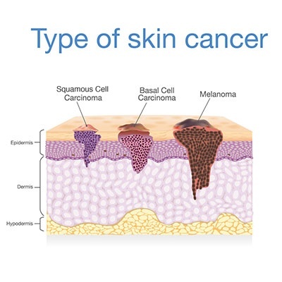
Skin cancer is becoming increasingly common and can spread to other organs. It is most often found in people over 40 and is more common in men than in women. Risk factors for skin cancer include:
- Prolonged exposure to ultraviolet (UV) radiation from sunlight, a major cause of skin cancer. Because the skin is the organ most exposed to sunlight, people who work in the sun, play outdoor sports or sunbathe regularly are at higher risk of skin cancer.
- Long-term consumption of food contaminated with chemicals such as arsenic, which can cause skin disease and increase the risk of skin cancer.
- Warts, moles, birthmarks or chronic wounds that remain irritated for a long time, which may develop into skin cancer.
- Smoking
- Fair skin
- A family history of skin cancer
Skin cancer can appear in several ways, for example moles, birthmarks or warts that increase in number or grow rapidly and change color. Such moles tend to have poorly defined borders and uneven color. Other signs include a smooth brown or black bump surrounded by a shiny, raised, rolled edge; an inflamed red rash; a chronic wound that does not heal or that bleeds; or a rough, uneven, very dark patch with poorly defined borders in an area that has never been injured or wounded. Skin cancer is most often found on the face, nose, head, arms, legs, palms, soles and trunk.

Human Epidermoid Carcinoma (A431)
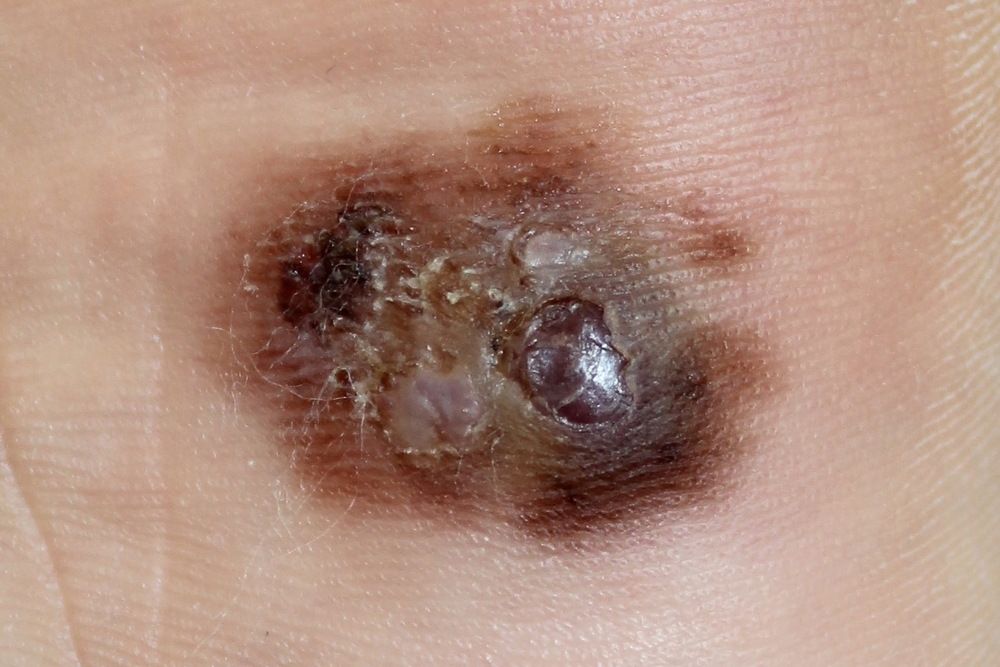
Epidermoid carcinoma, studied using the human A431 cell line, is a type of non-melanoma skin cancer that is less aggressive and has a lower mortality rate than malignant melanoma. Many studies have used A431 human epidermoid carcinoma cells to test inhibitory activity against skin cancer cells in vitro, in microplates (well plates) in which the cancer cells are grown in two- or three-dimensional culture (2D or 3D cell culture) in culture medium.
Testing on A431 Human Epidermoid Carcinoma Cells by Cell Culture
Herbal products, herbal extracts, dietary supplements and drugs are being developed for a range of health purposes, and their efficacy can be tested at the cellular level, including cytotoxicity to cancer cells, inhibitory activity and the effective concentration for inhibiting human epidermoid carcinoma (A431) skin cancer cells. In cell culture, cells or tissue obtained directly from living organisms are grown under controlled conditions so that the cancer cells grow, divide and multiply.
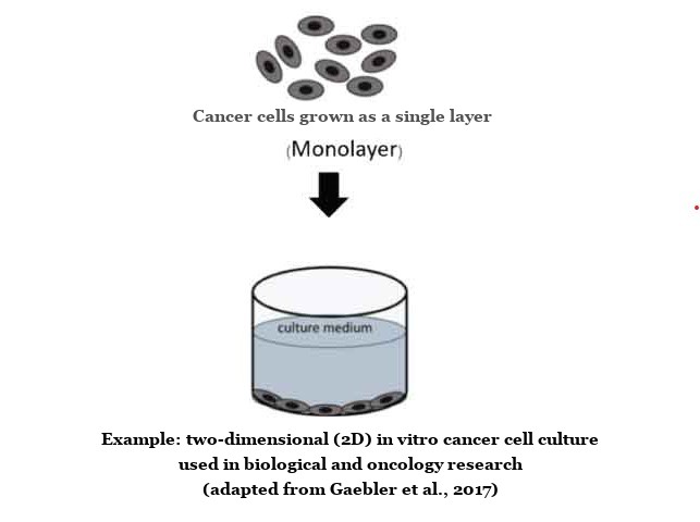
Cultured cancer cells share key characteristics with cancer cells transplanted into laboratory animals and with cancer cells in patients, so cell-based studies are carried out before testing in laboratory animals or clinical trials.
A431 Cell Inhibition Testing by MTT Assay
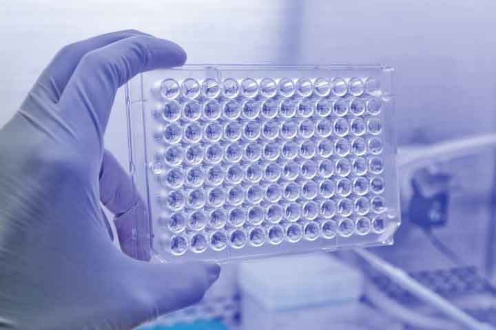
Inhibitory activity against human epidermoid carcinoma (A431) skin cancer cells grown in cell culture in microplates (well plates) is tested with the 3-[4, 5-Dimethylthiazol-2-yl]-2,5-diphenyltetrazolium bromide (MTT) assay. The MTT assay measures the inhibition of cancer cells in microplates based on the activity of mitochondrial dehydrogenase enzymes, which convert the yellow MTT (3-[4, 5-Dimethylthiazol-2-yl]-2,5-diphenyltetrazolium bromide) into purple formazan crystals.
Formazan crystals are therefore used as an indicator of cell viability: dead cancer cells remain clear and colorless, whereas living cancer cells form purple crystals inside the cell. When the crystals are dissolved in a solvent such as DMSO, they give a purple-blue solution whose absorbance can be measured with a spectrophotometer and is directly proportional to the number of living cells. The percentage survival of the cancer cells is then calculated and compared with a reference standard. Results can be reported as % inhibition of cancer cells or as the concentration of the substance that inhibits cancer cells by 50% (IC50).
Literature:
- สถาบันมะเร็งแห่งชาติ กรมการแพทย์ กระทรวงสาธารณสุข (in Thai)
- https://www.nci.go.th/th/Knowledge/skin.html
- ศูนย์เลเซอร์ผิวหนังและศัลยกรรมผิวหนัง ภาควิชาตจวิทยา คณะแพทยศาสตร์ศิริราชพยาบาล (in Thai)
- https://www.si.mahidol.ac.th/siriraj_online/thai_version/Health_detail.asp?id=1008
- โรงพยาบาลจุฬาลงกรณ์ สภากาชาดไทย, อาการต้องสงสัย อาจเป็นโรคมะเร็งผิวหนังเบซัลเซลล์ (in Thai)
- แสงสิริ อยู่อำไพ, ความเป็นพิษและกลไกการเหนี่ยวนำการตายแบบ apoptosis โดยสารสกัดจากเปลือกมังคุด (Garcinia mangostana) ในเซลล์มะเร็งผิวหนังชนิด human epidermoid carcinoma, 2555. (in Thai)
- Prin Sungwan, Wunchana Seubwai, Three-dimensional Cell Culture Model for In-Vitro Drug-Testing Platforms in Cancer Research, Srinagarind Med J 2021; 36(5): 625-633
- Gaebler M, Silvestri A, Haybaeck J, Reichardt P, Lowery CD, Stancato LF, et al. Three-dimensional patient-derived In vitro sarcoma models: promising tools for improving clinical tumor management. Front Oncol 2017; 7: 203.