
Service information about Wound Healing Efficacy Testing or Fibroblast Cell Generation
VISBIO Co., Ltd. offers analysis and testing services for wound healing efficacy or fibroblast cell generation conducted under 2D cell culture, applicable to various products in the health and beauty industry.
Wound healing efficacy testing is a process used to evaluate the body’s ability to recover damaged or injured tissues, such as those from accidents. The testing assesses the efficiency of fibroblast cell generation within the skin layer under 2D cell culture conditions. This testing is suitable for raw materials, herbal extracts, cosmetics, and dietary supplements with properties aimed at promoting skin recovery and healing and wound healing, such as plant-derived compounds like terpenes, terpenoids, carotenoids, and carotenes.
The Wound Healing Process
The skin serves a crucial role as a protective barrier against external factors, including physical hazards, pathogens, and Dehydration (loss of body fluids). It consists of two main layers: the epidermis and the dermis. When the skin sustains an injury, it immediately initiates a healing process to preserve its protective function.
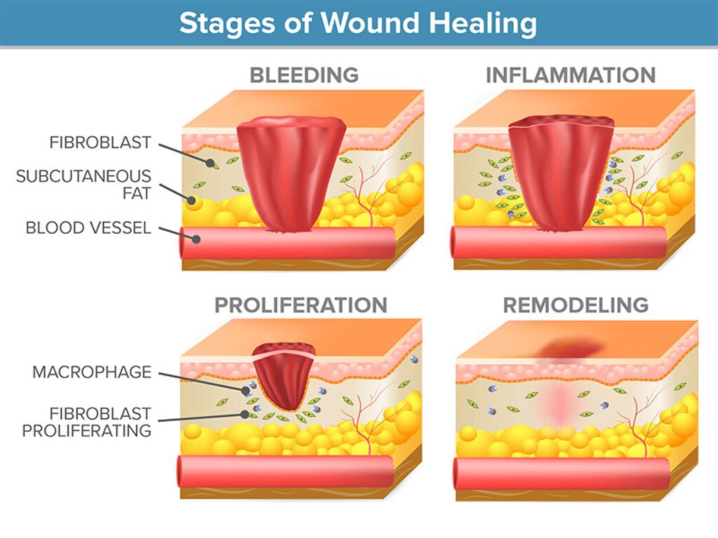
This wound healing or injury recovery process involves 3 distinct phases. In the first phase, known as the inflammation phase, the response begins within seconds after the injury occurs. Neutrophils and macrophages play a pivotal role in this phase by eliminating pathogens, releasing cytokines, and promoting the production of growth factors. These actions aid in the synthesis of collagen and other proteins, contributing to the formation of new tissue. The second phase, referred to as the proliferation or granulation phase, typically takes place 4-12 days following the injury. During this stage, fibroblasts are primarily responsible for generating collagen, leading to wound contraction and the development of new blood vessels. This phase is triggered by various cytokines, including TNF, TGF-B, and Vascular Endothelial Growth Factor (VEGF). In the final phase, called the remodeling or maturation phase, the collagen within the wound undergoes rearrangement and remodeling. Matrix metalloproteinases (MMP) may degrade collagen during this phase until a balance is achieved, ultimately resulting in the restoration and healing of the injured area.
The group of substances that promote wound healing
Substances that aid in the wound healing process can be categorized as follows:
- Volatile Oils: These belong to the secondary vegetal metabolism group and may possess properties such as anti-microbial, anti-inflammatory, and anti-oxidant effects.
- Plant-Derived Compounds: This category includes substances like terpenes, terpenoids, carotenoids, and carotenes. These compounds have anti-microbial properties and can inhibit the production of nucleic acids, such as robinetin, myricetin, and epigallocatechin. They can also disrupt cytoplasmic membranes and inhibit energetic metabolism. Examples of these compounds include BHA, p-coumaric acid, and caffeic acid, which hinder the arachidonic acid pathway.
- Propolis: Propolis has anti-inflammatory and anti-oxidant properties. Substances within this group are used in cosmetics and dietary supplements. Examples include essential oils, calendula officinalis, aloe vera, curcumin, musa sapientum (ripe banana), rubus ellipticus, allium ascalonicum linn (shallots), polygonum aviculare L (knotweed), and moringa oleifera.
Method for testing wound healing activity.
- In vitro Model
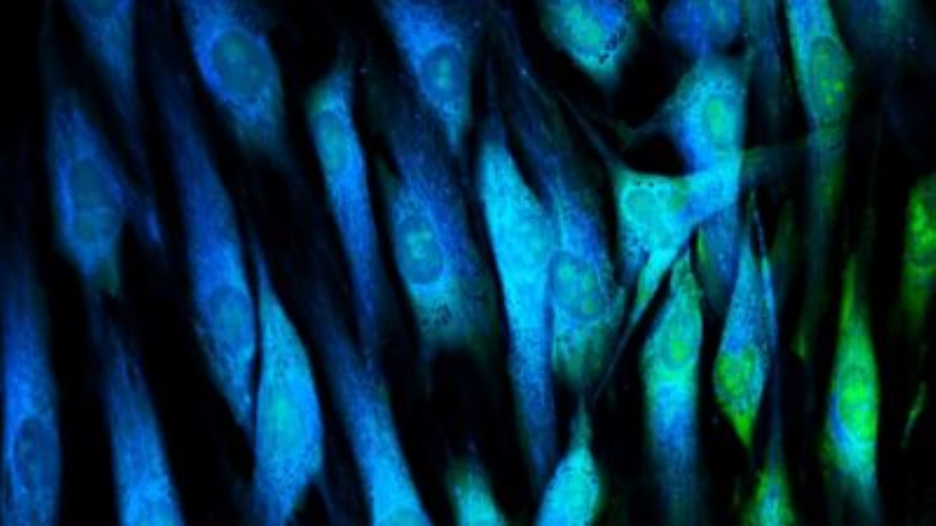
– Cell Migration (Chemotaxis) Testing: Culturing 2D cells in 96-well plates and then culturing them in various concentration conditions. Cell movement is observed at different time intervals using a microscope.
– Proliferation (Mitogenesis) Testing: Culturing cells to generate continuous fiber strands in 96-well plates, followed by incubation with various concentration samples to study cell division.
– Extracellular Matrix (ECM) Production: Fibroblast cells from the 96-well plate culture are treated with test samples after providing cell culture medium to achieve different concentrations. The cell culture medium is then analyzed to quantify the production of fibroblast ECM and is compared to the control group with no test samples. - Animal studies model: This includes various animal models such as Granuloma Models, Incision Models, Excision Models, Open-Wound Models, and Burn Models.
- Impaired Healing Model: Testing of chronic wounds, including those arising from complications related to diabetes, such as diabetic foot ulcers.
- Tissue Repair in Humans
Literature:
- Cañedo-Dorantes, Luis, and Mara Cañedo-Ayala. “Skin acute wound healing: a comprehensive review.” International journal of inflammation 2019 (2019).
- Greenhalgh, David G. “Models of wound healing.” Journal of Burn Care & Rehabilitation 26.4 (2005): 293-305.
- Georgescu, Mihaela, et al. “Natural compounds for wound healing.” Worldwide Wound Healing—Innovation in Natural and Conventional Methods. Portugal: IntechOpen (2016): 61-89.
- Nayak, B. S., and Lexley M. Pinto Pereira. “Catharanthus roseus flower extract has wound-healing activity in Sprague Dawley rats.” BMC Complementary and Alternative medicine 6.1 (2006): 1-6.