
Service information on Testing the Inhibition Properties of T-cell Leukemia White Blood Cells
VISBIO Co., Ltd. provides services for testing and analyzing the inhibition properties of T-cell Leukemia white blood cancer cells in a laboratory setting. This testing involves culturing cancer cells in a 2D cell culture and is suitable for in-depth research with the objective of developing herbs that can inhibit cancer cell growth.
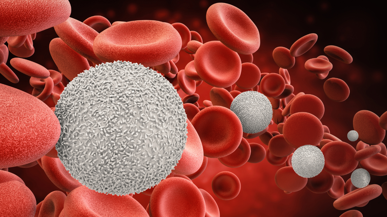
The rate of death in the population caused by white blood cell leukemia is 4 per 100,000 people. Leukemia is a type of cancer within the blood system that originates from abnormal growth of immature white blood cells in the bone marrow. This leads to an abnormal increase in white blood cells without control, resulting in an accumulation of white blood cells in the bone marrow and their release into the bloodstream. T-cell Leukemia (Jurkat) is one type of white blood cell cancer derived from humans and is commonly used to study the biology of leukemia, exploring the inhibition of leukemia cells, both externally and in Well Plate, under 2D or 3D cell culture conditions, and evaluating anti-cancer products in cell nutrition.
Get to know Leukemia
Information from the Strategy, Ministry of Public Health, 2017-2021, revealed that the mortality rate of leukemia is 4 per 100,000 people. Leukemia is one type of cancer within the blood system that originates from the abnormal growth of immature white blood cells in the bone marrow. This leads to an abnormal increase in white blood cells that cannot be controlled. As a result, white blood cells accumulate excessively in the bone marrow and are released into the bloodstream, leading to abnormally high levels of white blood cells in the bloodstream.

Leukemia is divided into two major groups:
- Acute Lymphocytic Leukemia (ALL): This type of leukemia results from an increased number of immature white blood cells that cannot grow into mature white blood cells. This rapid increase in immature white blood cells can lead to severe and rapid symptoms, including weakness, fatigue, pale skin due to anemia, fever, and susceptibility to infections. This is because mature white blood cells, which are responsible for infection defense, decrease, and the risk of bleeding increases. It may present with skin hemorrhages, especially in various parts of the body. In most cases, ALL has more severe symptoms compared to the chronic type.
- Chronic Lymphocytic Leukemia (CLL): This type results from an abnormal increase in white blood cells, which can still develop into mature white blood cells. Symptoms may be mild or absent initially. However, the majority of patients do not exhibit symptoms early on. As the level of white blood cells in the bloodstream increases, symptoms like fatigue, anemia, and swollen lymph nodes may occur. The spleen may also enlarge, leading to abdominal distension or discomfort, especially in the upper left abdomen.
The exact causes of leukemia remain unclear. However, epidemiological studies have identified some factors that may contribute to the risk of developing leukemia. These include genetics, with a higher incidence of the disease found in North America and Western Europe, and a lower incidence in Asia and Latin America. Other risk factors may include exposure to radiation, as studies of Japanese survivors of the atomic bombings during World War II have shown a much higher risk of developing leukemia. Additionally, exposure to certain chemicals or substances, such as those found in tanneries, dyes, pesticides, hair dyes, or sprays, may also increase the risk. Finally, prior chemotherapy for another cancer is associated with an increased risk of developing leukemia, particularly 2-10 years after chemotherapy.
Leukemia type T-cell Leukemia (Jurkat)
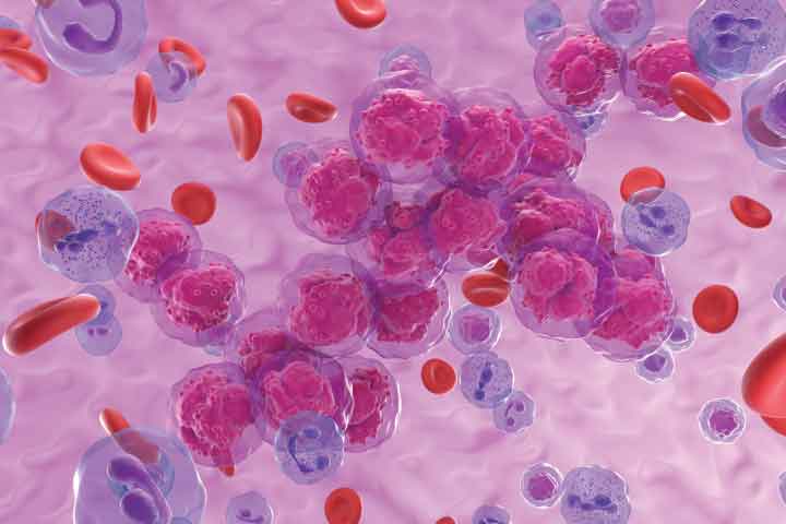
T-cell Leukemia (Jurkat) is a type of white blood cell cancer that originates in humans. It is one of the white blood cell cancer cells commonly used for studying the biology of leukemia. Research focuses on understanding how these leukemia cells inhibit the growth of cancer cells either outside or within Well Plate under two-dimensional or three-dimensional conditions (2D or 3D Cell Culture – Anti-cancer) with the aid of cell culture medium.
Testing T-cell Leukemia (Jurkat) White Blood Cell Cancer Cells in Cancer Cell Culture
Currently, there is the development of herbal products, plant extracts, dietary supplements, or medications designed for nourishment and anti-inflammation. These products can be tested at the cellular level regarding their toxicity to cancer cells, inhibitory properties, or appropriate concentration for inhibiting T-cell Leukemia (Jurkat) white blood cell cancer cells.
The advanced testing process involves taking cells or tissue directly from living organisms and cultivating them under controllable environmental conditions to promote the growth, division, or multiplication of cancer cells. This is similar to culturing cancer cells taken from animal experiments or cancer cells within a patient’s body. It is a preliminary test before proceeding to animal experiments or further clinical trials.

Figure: Demonstrating an example of two-dimensional cell culture in an in-vitro setting used for studying cell biology and cancer research (Gaebler et al., 2017).
Testing the Inhibition Efficiency of T-cell Leukemia (Jurkat) White Blood Cell Cancer Cell Inhibition using the MTT Assay
The inhibition efficiency of T-cell Leukemia (Jurkat) white blood cell cancer cells, cultivated at the cellular level or in microplates (Well Plate), is tested using the Methyl tetrazolium 3-[4,5-Dimethylthiazol-2-yl]-2,5-diphenyltetrazolium bromide (MTT) assay.
The MTT assay is a method for testing the inhibitory effects on cancer cells in microplates based on the ability of Dehydrogenase enzymes in mitochondria to work. The yellow MTT (3-[4,5-Dimethylthiazol-2-yl]-2,5-Diphenyltetrazolium Bromide) dye is converted into a purple Formazan pigment.
In this assay, non-viable cancer cells appear transparent without color, while viable cancer cells display purple formazan within the cells. When dissolved in a solubilizing solution like DMSO, it produces a blue-purple solution that can be quantified for light absorbance using a spectrophotometer. This measurement correlates directly with the quantity of viable cells. The results can be reported as a percentage of cancer cell inhibition or as the concentration required to inhibit 50% of the cells (IC50).
Literature:
- ลลิตา นรเศรษฐ์ธาดา, ลิวคีเมีย, หน่วยโลหิตวิทยา ภาควิชาอายุรศาสตร์ คณะแพทยศาสตร์ มหาวิทยาลัยเชียงใหม่, สมาคมโลหิตวิทยาแห่งประเทศไทย
- มะเร็งเม็ดเลือดขาว รู้ทันป้องกันได้, โรงพยาบาลราชวิถี
- นพดล ศิริธนารัตนกุล, โรงมะเร็งเม็ดเลือดขาว, ภาควิชาอายุรศาสตร์ คณะแพทยศาสตร์ศิริราชพยาบาล
- โรคมะเร็งเม็ดเลือดขาว, สถิติสุขภาพคนไทย, กองยุทธศาสตร์และแผนงาน กระทรวงสาธารณสุข, 2560-2564
- Gaebler M, Silvestri A, Haybaeck J, Reichardt P, Lowery CD, Stancato LF, et al. Three-dimensional patient-derived In vitro sarcoma models: promising tools for improving clinical tumor management. Front Oncol 2017; 7: 203.