

Service information on testing the inhibitory efficacy of prostate cancer cells line (PC-3)
VISBIO Co., Ltd. offers testing and analysis services for the inhibitory efficacy of prostate cancer cells line (PC-3) in a laboratory setting. Our team of scientific experts conducts tests by culturing cancer cells in a 2D cell culture environment. This testing is suitable for in-depth research aimed at developing herbal products that can effectively inhibit prostate cancer cells. The prostate is an organ in the male reproductive system located below the bladder and in front of the rectum. It generally increases in size with age. According to data from the National Cancer Institute in 2021, prostate cancer ranks as the 5th most common cancer among Thai men, with a continuous upward trend. At present, various herbal products, herbal extracts, dietary supplements, or medications are being developed to support prostate health or counter inflammation in the prostate. These products can be tested at the cellular level regarding their cytotoxic effects, inhibitory properties, or determining the suitable concentration for inhibiting prostate cancer cells line (PC-3). If you are interested in analyzing the inhibitory efficacy of products against prostate cancer, please feel free to contact our company through any of the provided channels.
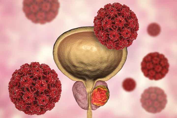
Get to know Prostate Cancer
The prostate is an organ in the male reproductive system located beneath the bladder and in front of the rectum. It’s about the size of a walnut and surrounds a portion of the urethra. Typically, the prostate gland enlarges with age, gradually causing constriction of the urethra or bladder neck, leading to urinary difficulties. This condition is known as benign prostatic hyperplasia (not cancer) and can be managed with medications or surgery.

According to information from the National Cancer Institute in 2021, prostate cancer is the fifth most common cancer among Thai men, and there’s a continuous upward trend in its prevalence. Prostate cancer develops when cells or tissues within the prostate gland grow and divide abnormally and rapidly or cannot control cell division. This leads to the obstruction of the urethra, destruction of normal prostate cells, and potential spread to other vital organs such as the liver, kidneys, lungs, and bones.
Prostate Cancer Cells Line (PC-3)

Prostate cancer cells line (PC-3) are human-derived prostate cancer cells. They are a commonly used type of prostate cancer cells in the study of prostate cancer biology. The research involves investigating their anti-cancer properties in both two-dimensional (2D) and three-dimensional (3D) cell cultures.
Testing Prostate Cancer (PC-3) in Prostate Cancer Cell Culture (Cell Culture - Anti-cancer)
Currently, there is a significant development in the development of herbal products, extracts from herbs, nutritional supplements, as well as medicines designed to nourish or combat inflammation in the prostate. These products are formulated to be tested for their effectiveness at the cellular level, assessing its toxicity towards cancer cells, inhibitory properties, or the appropriate concentration required to inhibit prostate cancer cells of the Prostate cancer cells line (PC-3).
The advanced testing process involves taking cells or tissue directly from living organisms and cultivating them under controllable environmental conditions to promote the growth, division, or multiplication of cancer cells. This is similar to culturing cancer cells taken from animal experiments or cancer cells within a patient’s body. It is a preliminary test before proceeding to animal experiments or further clinical trials.

Figure: Demonstrating an example of two-dimensional cell culture in an in-vitro setting used for studying cell biology and cancer research (Gaebler et al., 2017).
Testing the inhibitory effects on Prostate cancer cells line (PC-3) using MTT assay
The MTT assay is employed to assess the inhibitory properties of compounds on cancer cells grown in microplates. This assay relies on the functionality of dehydrogenase enzymes within the mitochondria, which convert the yellow MTT (3-[4, 5-Dimethylthiazol-2-yl]-2,5-Diphenyltetrazolium Bromide) into purple formazan. Therefore, formazan can be used to indicate cell viability.
In this assay, non-viable cancer cells appear transparent without color, while viable cancer cells display purple formazan within the cells. When dissolved in a solubilizing solution like DMSO, it produces a blue-purple solution that can be quantified for light absorbance using a spectrophotometer. This measurement correlates directly with the quantity of viable cells. The results can be reported as a percentage of cancer cell inhibition or as the concentration required to inhibit 50% of the cells (IC50).
Literature:
- มะเร็งต่อมลูกหมาก ตรวจพบเร็ว หายขาดได้, โรงพยาบาลศิริราช ปิยะมหาราชการุณย์
- โรคมะเร็งต่อมลูกหมาก, คณะแพทยศาสตร์โรงพยาบาลรามาธิบดี มหาวิทยาลัยมหิดล ศูนย์ความเป็นเลิศด้านโรคมะเร็ง
- แผนยุทธศาสตร์สถาบันมะเร็งแห่งชาติ, สถาบันมะเร็งแห่งชาติ กรมการแพทย์ กระทรวงสาธารณสุข, พ.ศ. 2562 – 2565
- ทะเบียนมะเร็งระดับโรงพยาบาล, สถาบันมะเร็งแห่งชาติ กรมการแพทย์ กระทรวงสาธารณสุข, พ.ศ.2564
- Gaebler M, Silvestri A, Haybaeck J, Reichardt P, Lowery CD, Stancato LF, et al. Three-dimensional patient-derived In vitro sarcoma models: promising tools for improving clinical tumor management. Front Oncol 2017; 7: 203.